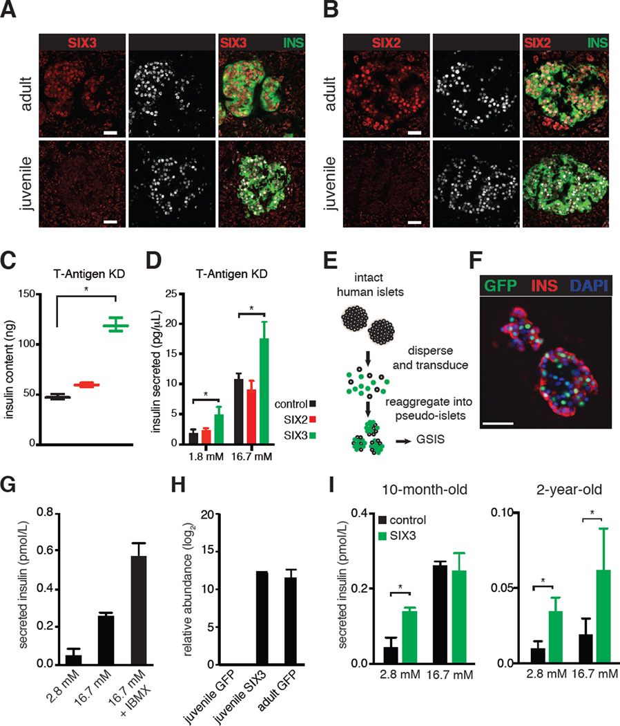
Figure 5. SIX3 and SIX2 increase with age specifically in human β-cells and enhance β-cell maturation.
SIX3 (A) and SIX2 (B) immunostaining of adult (31-year-old) and juvenile (4-year-old) human pancreas sections, scale bar: 100 µm. Insulin content (C) and secreted insulin levels (D) of EndoC-βH1 cells expressing GFP, SIX2 or SIX3 without T-Antigen (T-Antigen KD). Asterisks indicate statistically significant results reproduced in multiple experimental replicates (P < 0.05, two-way ANOVA followed by Fisher’s Least Significant Difference test). Bars indicate S.D. (E) Schematic detailing pseudo-islet techniques. Human islet cells are enzymatically dispersed, and transduced with lentivirus which co-expresses a GFP transgene. 5–7 days after dispersion, islet cells spontaneously re-aggregate into pseudo-islet clusters can be assayed using glucose-stimulated insulin secretion (GSIS) in vitro. (F) Representative immunohistologic images of human pseudo-islets stained with Insulin, GFP or DAPI, scale bar: 50 µm. (G) Bar graphs show secreted insulin levels of cultured human pseudo-islets exposed to glucose or glucose +IBMX. Secreted insulin is normalized to total insulin content. (H) Bar graphs indicate relative abundance of SIX3 transcript detected in pseudo-islets after transduction with GFP or SIX3 lentiviral vectors. (I) Graphs show secreted insulin levels from cultured pseudo-islets obtained from juvenile donors, expressing GFP alone (control) or SIX3. Secreted insulin is normalized to total insulin content (* P < 0.05, t-test). Error bars indicate S.D.

