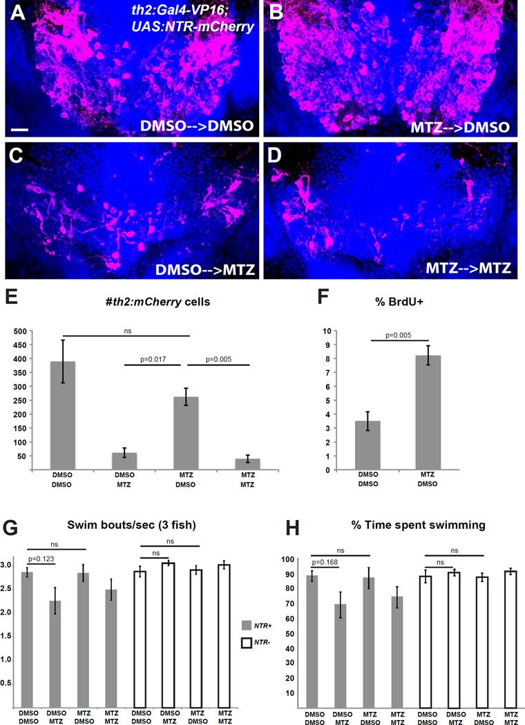
Figure 4. th2+ cells are replaced after ablation and mediate functional recovery of swimming behavior.
(A–D) Representative brains from th2:Gal4-VP16; UAS:NTR-mCherry larvae that were treated with 0.5% DMSO or 5mM MTZ from 5–7 dpf, followed by 10 mM BrdU from 8–10 dpf and allowed to recover for two weeks (10–25 dpf) before acute treatments with 0.5% DMSO or 5mM MTZ. (E) Recovery of th2+ cells measured by total cell number. Analysis of the 4 groups by 2-way ANOVA indicates that the 25 dpf treatment has a significant effect on the number of th2:mCherry cells at 28 dpf (p=0.0001), while the 7 dpf treatment does not (p=0.082); nor is there a significant interaction between two successive treatments (p=0.20). Adjusted p values shown for pairwise comparisons are based on Bonferroni Multiple Comparison test. n=3 brains for larvae untreated at 7 dpf, n=4 brains for larvae treated with MTZ at 7 dpf. (F) Percent labeled by BrdU at 8 dpf. Error bars=SEM, n=5 brains. p value based on Student t-test. (G–H) Recovery of behavior 3 weeks after ablation as measured by swimming frequency (G) and time spent swimming (H), n=5 groups for each condition except n=4 for wild-type larvae treated with MTZ+DMSO after exclusion of an outlier (p<0.001, Grubbs’ test). Statistical analyses were performed separately for the wild-type and NTR+ larvae. Two-way ANOVA of wild-type larvae shows no significant effect of either the 7 dpf treatment (p=0.95 for swim frequency; p=0.89 for swim time) or the 25 dpf treatment (p=0.13 for swim frequency; p=0.37 for swim time) at 28 dpf, and there is no significant interaction between the successive treatments (p=0.66 for swim frequency; p=0.76 for swim time). For NTR+ larvae, 2-way ANOVA indicates that the 7 dpf treatment does not affect swimming behavior at 28 dpf (p=0.60 for swim frequency; p=0.80 for swim time), while the 25 dpf treatment has a significant effect (p=0.034 for swim frequency; p=0.032 for swim time). There was no significant interaction between the successive treatments (p=0.55 for swim frequency; p=0.64 for swim time). Adjusted p values shown for pairwise comparisons are based on Bonferroni Multiple Comparison test. Images in (A–D) are ventral maximum intensity confocal Z-projections of the hypothalamic posterior recess. Scale bar = 10µM. See Figure S2 for representative plots of swimming behavior in ablated and unablated animals.

