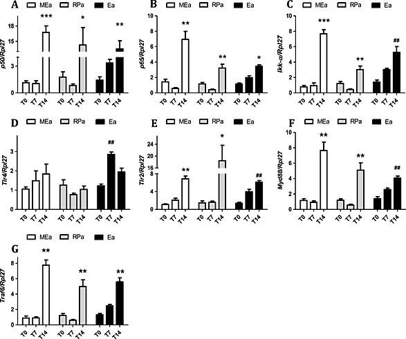
Figure 3.

Gene expression of transcription factors involved in pro‐inflammatory pathways in isolated adipocytes along cachexia progression. Results are expressed as mean ± standard error of the mean (n = 4). Adipocytes isolated from adipose tissue were obtained from animals 0, 7, and 14 days after inoculation of tumour cells: *P < 0.01 vs. all the groups for the same tissue; # P < 0.05 vs. T0; **P < 0.01 vs. all the groups for the same tissue; ## P < 0.01 vs. T0. Mesenteric adipocytes (MEa), retroperitoneal adipocytes (RPa), and epididymal adipocytes (Ea). (A) gene expression of p50, (B) gene expression of p65, (C) gene expression of Ikk‐α, (D) gene expression of TLR4, (E) gene expression of TLR2, (F) gene expression of Myd88, and (G) gene expression of Traf6.
