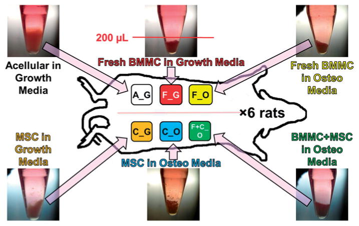
Figure 1.

Schematic of microbead implants in rat subcutaneous dorsum. Microbeads cultured for a total of 20 days (n = 6, shown in photos of centrifuge tube cultures) were subsequently mixed with 500 μl of fibrin gel carrier and injected subcutaneously in rat dorsum. Microbead implant types, according to type of encapsulated cells and culture media, were A_G, F_G, F_O, C_G, C_O, and F+C_O, where A = acellular; F = freshly isolated BMMC; C = culture-expanded MSC; G = growth medium, O = osteogenic medium. Ectopic implants were harvested at 5-weeks, and analyzed by microCT and histology.
