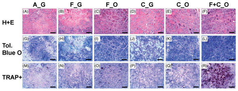
Figure 5.

Histology of 5-week ectopic microbead implants. Representative views of sections (7 μm) of microbead implants stained with Hematoxylin and Eosin (H + E), Toluidine Blue O (Tol. Blue O), or for tartrate-resistant acid phosphatase positive cells (TRAP+). Microbead types of each implant, according to type of encapsulated cells and culture media, were A_G, F_G, F_O, C_G, C_O, and F + C_O, where A = acellular; F = freshly isolated BMMC; C = culture-expanded MSC; G = growth medium, O = osteogenic medium. Scale bar = 200 μm. Images best viewed in color.
