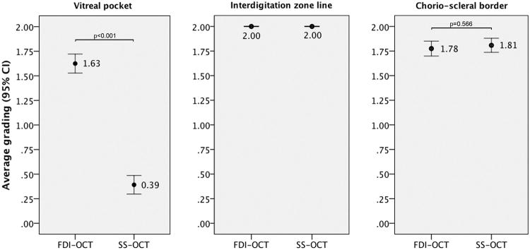
Figure 1.
Qualitative comparison of the FDI SD-OCT imaging to the SS-OCT imaging with respect to visualization of the anterior border of the premacular bursa, interdigitation zone line, and chorio-scleral boundary. The grading system was the following: grade 0 indicated that the structure was not seen, grade 1 indicated that the structure was barely seen, and grade 2 indicated that the structure was clearly seen. The scores were added for the two graders, who were masked to OCT imaging modality. Shown are average scores for each OCT modality with 95% confidence intervals.

