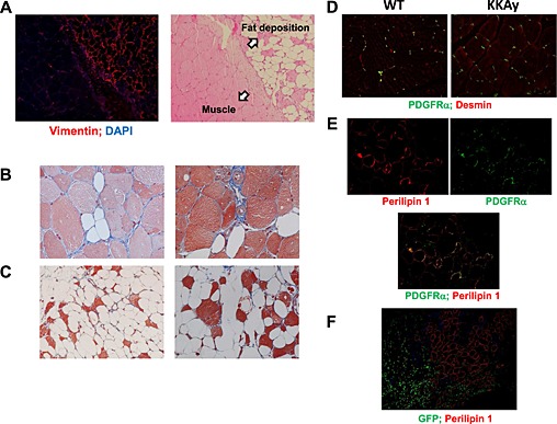
Figure 5.

Origin of fat formation after cardiotoxin treatment in KKAy. (A) Immunofluorescent staining with anti‐vimentin antibody at the border between fat formation and muscle of 26‐week‐old mice at ×100 magnification. Masson's trichrome staining of fat formation in KKAy 2 weeks after cardiotoxin treatment of 8‐week‐old (B) and 26‐week‐old (C) mice at ×200 magnification. (D) Immunofluorescent staining with anti‐platelet‐derived growth factor (PDGF) receptor alpha and anti‐desmin antibodies in non‐injured muscle at ×100 magnification. (E) Representative photos of immunofluorescent staining with anti‐perilipin 1 and anti‐PDGF receptor alpha antibodies at ×200 magnification. (F) Representative photos of immunofluorescent staining with anti‐perilipin 1 and anti‐GFP antibodies in green fluorescent protein (GFP)‐chimeric KKAy at ×100 magnification.
