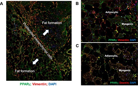
Figure 7.

(A) Immunofluorescent staining with anti‐vimentin and anti‐peroxisome proliferator‐activated receptor gamma (PPARγ) antibodies in border between fat formation and satellite cells in 26‐week‐old KKAy after cardiotoxin treatment. Blue line shows the border. High magnification (×400) view of immunofluorescent staining with anti‐vimentin and anti‐PPARγ antibodies (B) and anti‐desmin and anti‐PPARγ antibodies (C) in the border between fat formation and satellite cells in 26‐week‐old KKAy after cardiotoxin treatment. DAPI, 4',6‐diamidino‐2‐phenylindole.
