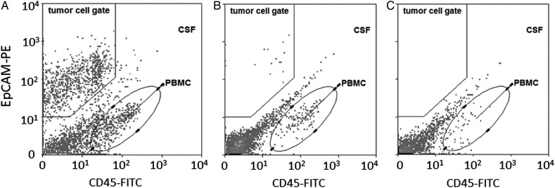
Fig. 2.
Examples of EpCAM-based flow cytometry plots in CSF in individual patients. (A) NSCLC patient with LM with EpCAM-positive CTCs (162 CTCs/mL); CSF cytology was positive (not shown). (B) Breast cancer patient with LM with EpCAM-positive CTCs (3 CTCs/mL). CSF cytology was negative (not shown). (C) Breast cancer patient without LM. No EpCAM-positive CTCs in CSF. CSF cytology was also negative (not shown).

