Fig. 3.
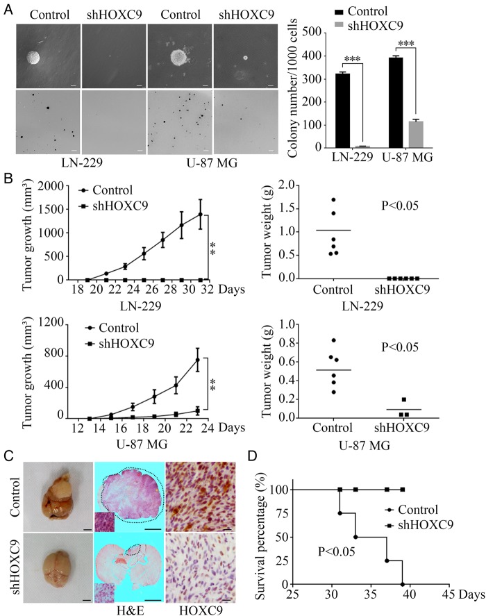
HOXC9 is required for glioblastoma cell self-renewal and tumorigenesis of glioblastoma cells. (A) Soft agar assays were performed after HOXC9 knockdown in LN-229 and U-87 MG cell lines. The quantification of colony numbers is also presented (error bars, SEM, n = 3; scale bars, upper = 50 μm, lower = 200 μm). (B) Subcutaneous xenograft tumor growth was monitored after HOXC9 knockdown in LN-229 and U-87 MG cells. Tumor volumes were measured using a caliper every 2 days (error bars, SEM, n = 6). Tumor weights are presented in scatterplots with horizontal lines indicating the mean. (C) Orthotopic implantation was performed after HOXC9 knockdown in U-87 MG cells. shGFP was used as the control. Representative images of the original tumor formation (left), hematoxylin and eosin(H&E) staining (middle), and immunohistochemistry analysis of HOXC9 expression (right) are presented. Scale bars: left and middle, 3 mm; right, 20 μm. (D) Survival rates were analyzed after orthotopic implantation of shHOXC9 U-87 MG cells. n = 4, P < .05 by the log-rank test for significance. Statistical analysis was performed using 2-tailed Student t tests, ** P < .01, *** P < .001.

