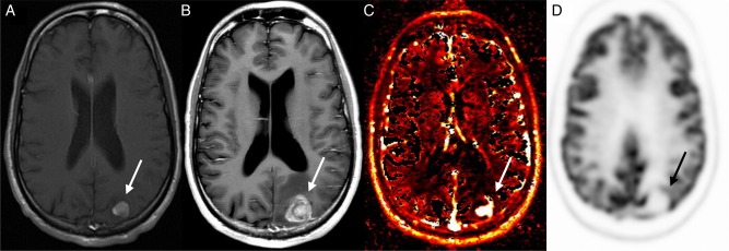
Fig. 2.
Patient example of tumor progression detected by DCE-MRI perfusion. Images obtained in a 30-year-old man with metastatic sarcoma who underwent SRS to a left parietal lobe metastasis. Axial contrast-enhanced T1-weighted image before treatment (A) shows an enhancing mass (arrow) that increases in size 6 months after treatment (B). Vp map of the enlarging mass (C) demonstrates increased perfusion; however, PET-CT showed no abnormal FDG uptake (D). Pathology confirmed progression.

