Figure 1.

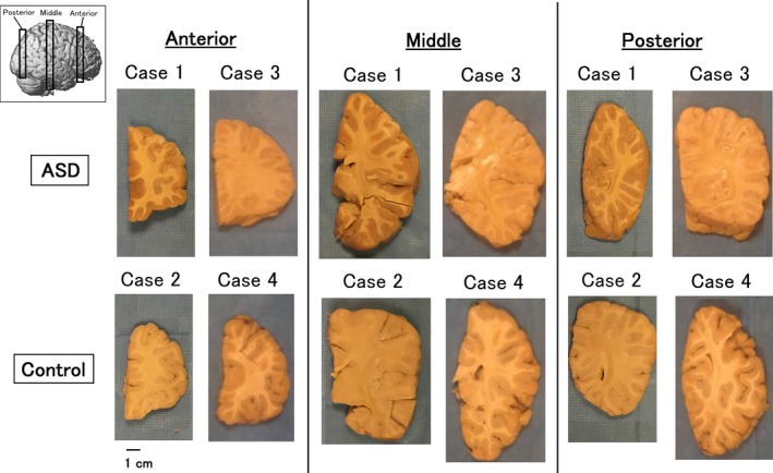
Brain specimens studied. See details in text. Approximate slice locations are shown in left upper corner (three‐dimensional brain image is from http://www.fil.ion.ucl.ac.uk/spm).

Brain specimens studied. See details in text. Approximate slice locations are shown in left upper corner (three‐dimensional brain image is from http://www.fil.ion.ucl.ac.uk/spm).