
Figure 5.
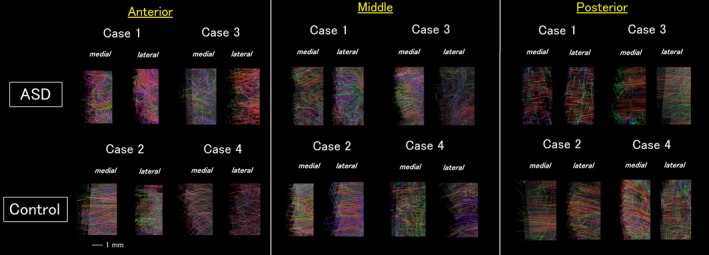
High angular resolution diffusion imaging tractography of the gray matter pathways. Both medial and lateral surfaces of the anterior slices showed clear disorganization in the Autism spectrum disorder brains. In the medial slices, the lateral surface showed more sparse, less coherent pathways compared to the controls. The posterior slices did not show clear difference between two groups. The color‐coding of tractography pathways was the one used in Figures 2 and 3.