
Figure 3. Structure of the RING dimers and interaction with the E2~Ub intermediate.
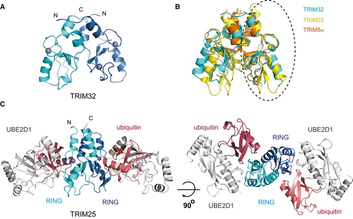
- Structure of the TRIM32 RING dimer in ribbon representation with each RING monomer coloured in cyan and blue and the Zn2+ ions as grey spheres.
- Overlap of the RING dimers of TRIM32 (cyan, 5FEY.pdb), TRIM25 (yellow, 5FER.pdb) and TRIM5α (orange, 4TKP.pdb). The structures were overlapped on the circled RING domain. This overlap shows that the structures of the RINGs are very similar but that there are differences in the relative orientations of the two RINGs.
- Structure of the TRIM25 RING/E2˜Ub complex with the RING domains in the same colour scheme as in (A), UBE2D1 in grey and the ubiquitin molecules in salmon and red.