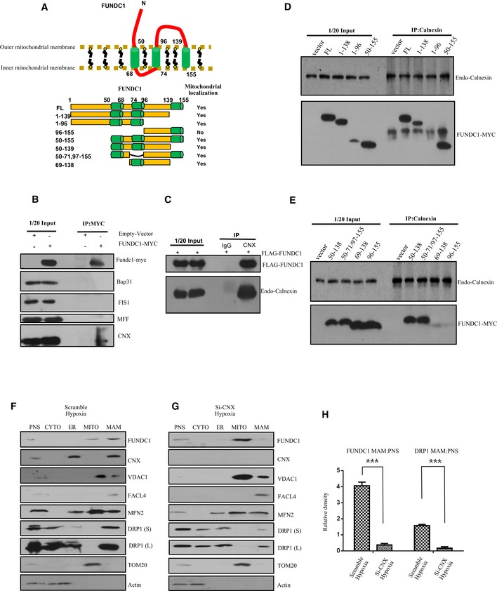
Figure 2. The ER membrane protein calnexin is required for the accumulation of FUNDC1 in the MAM .

-
ATop: Cartoon representing the topology of the FUNDC1 protein in the mitochondrial membrane. The cytoplasmic domain that interacts with the ER protein calnexin is shown. Bottom: Illustration of the different FUNDC1 truncation variants.
-
BHeLa cells were transfected with FUNDC1‐MYC or empty vector for 24 h. Cell lysates were immunoprecipitated by anti‐MYC antibody and immunoblotted with the indicated antibodies.
-
CHeLa cells were transfected with FLAG‐FUNDC1. About 24 h post‐transfection, cell lysates were immunoprecipitated by anti‐calnexin and immunoblotted with anti‐FLAG and anti‐calnexin antibodies.
-
D, EHeLa cells were transfected with the indicated FUNDC1‐MYC constructs for 24 h. Cell lysates were immunoprecipitated using anti‐calnexin and immunoblotted using anti‐MYC and anti‐calnexin antibodies.
-
FImmunoblots of subcellular fractions from HeLa cells transfected with scramble siRNA and exposed to hypoxia (1% O2) for 5 h. PNS: post‐nuclear supernatant; CYTO: cytosol; ER: endoplasmic reticulum; MITO: mitochondria; MAM: mitochondrial‐associated membrane. DRP1 (S) and DRP1 (L) indicate short and long exposures, respectively.
-
GImmunoblots of subcellular fractions from HeLa cells transfected with calnexin siRNA (siCNX) and exposed to hypoxia (1% O2) for 5 h. PNS: post‐nuclear supernatant; CYTO: cytosol; ER: endoplasmic reticulum; MITO: mitochondria; MAM: mitochondrial‐associated membrane. DRP1 (S) and DRP1 (L) indicate short and long exposures, respectively.
-
HQuantification of the MAM:PNS ratio of FUNDC1 or DRP1 in HeLa cells treated with scramble siRNA or siCNX and exposed to hypoxia (1% O2) for 5 h. Data are presented as mean ± s.e.m. from three independent experiments, ***P < 0.001.
Source data are available online for this figure.
