
Figure 3.
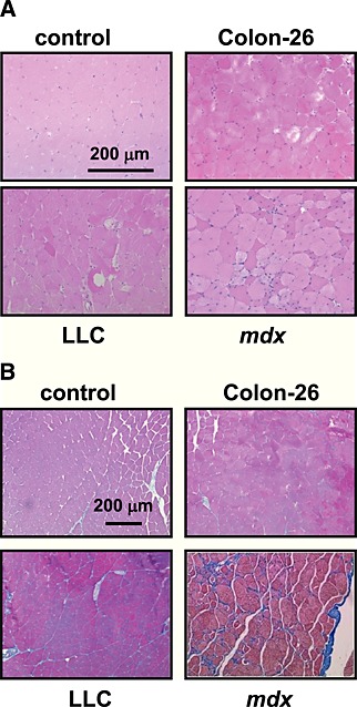
Histological features of cancer cachexia muscles. Representative images of haematoxylin and eosin staining (A) and Masson's trichrome staining (B) of gastrocnemius muscles sections from cancer‐bearing mice or mdx mice. Scale bar, 200 µm. Although C57BL/6 data were represented as control, a similar result was obtained from other control strain (CD2F1). LLC, Lewis lung carcinoma.