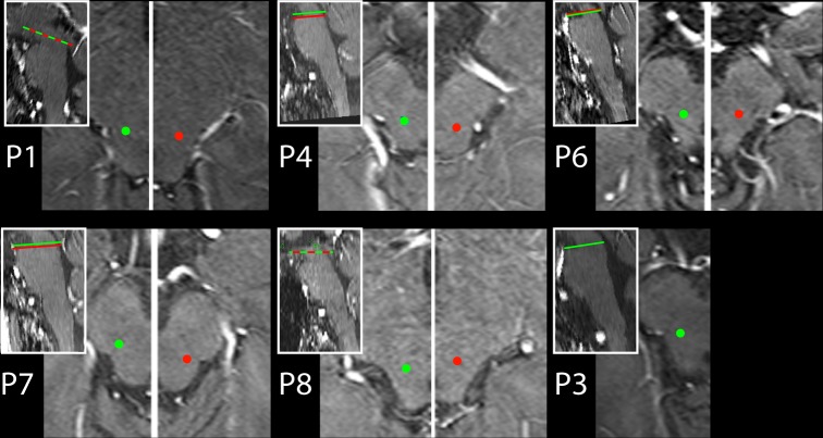
Fig 2. DBS electrode contact locations.
The location of the active electrode contacts in the right (green dots) and left (red dots) PPN region. Shown are axial MRI transsections in each patient. Left and right side transsections display different planes along the rostro-caudal axis in P4, P6, and P7. Respective planes are highlighted by green (right side) and red lines (left side) in the inserts showing midline sagittal transsections.

