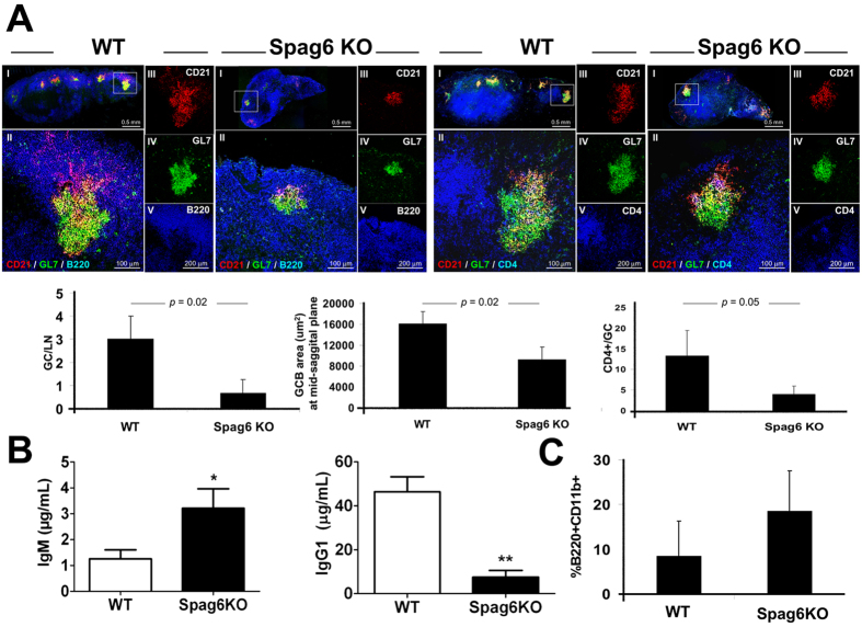
Figure 4. Defective humoral immune response in Spag6KO mice.
(A) Reduced GC formation and GC CD4+ T cells in draining LN (day 14) of WT and Spag6KO reconstituted mice. (I) Low magnification image showing the whole LN at a mid saggital section. The GC in white box is shown at higher magnification in (II) and Separate channel recordings (III, IV, V) are provided to the right of the overlay of the presented GC. The left panel shows FDCs (CD21+, red), GCB cells (GL7+, green), and B220/CD45R (pan B cell marker, blue) in the lymph node cortex. The right panel shows FDCs (CD21+, red), GCB cells (GL7+, green), and CD4+ T cells including GC CD4+ T cells (blue). Morphometric analysis of GCs illustrated in histograms shows reduced number and size of GCs and CD4+ T cells per GC in Spag6KO. (B) Day 14 NP-KLH specific (B, left) IgM and (B, right) IgG1 levels in sera. (C) Percentage of peritoneal B1 B cells (B220+ CD11b+) collected at day 14 post-immunization. N = 4 − 6 per group, 3 independent experiments. *p < 0.05, **p < 0.005.

