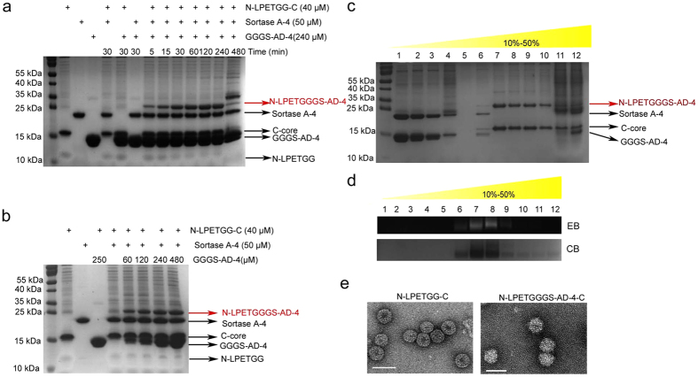
Figure 2. Chemoenzymatic coupling of GGGS-AD-4 to N-LPETGG-C VLPs without impairing the VLP structure.
(a) Time-dependent assay of sortagging GGGS-AD-4 to VLPs. Sortagging reached a balance after 1 hour of incubation. Target product was labeled with a red arrow. Other components used in coupling system were instructed in black arrows. (b) Optimization of the concentrations of the substrate. No apparent increase in the target product was observed when the substrate was more than 3-fold. (c) Tricine-SDS-PAGE analysis and (d) NAGE analysis were performed to monitor the sucrose gradient density ultracentrifugation separation of the target product. NAGE was stained with ethidium bromide (EB) and coomassie brilliant blue (CB). (e) VLP structures were confirmed by transmission electron microscopy (TEM). White bar: 50 nm.

