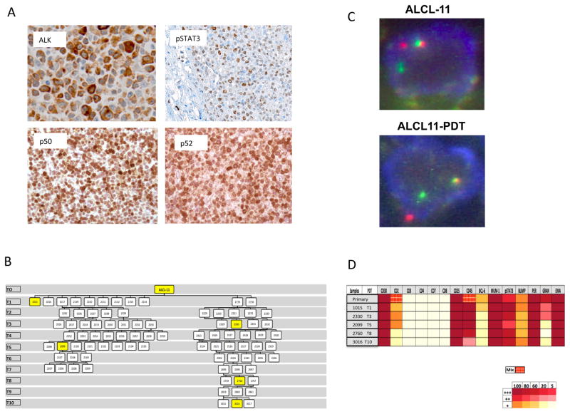
Fig 1. The ALCL-11 Patient Derived Tumorgraft line mimics its corresponding primary tissues sample.
(A) Immunohistochemistry stains performed on primary diagnostic sample (ALCL-11) show a strong ALK cytoplasmic staining with an antibody (D5F3, Cell Signaling Technology) specific for an intracytoplasmic peptide of the human ALK gene (x 400). Tissue sections stained with specific antibody against human phospoSTAT3 (pSTAT3, x100), p50 and p52 demonstrate a variable number lymphoma cells with nuclear positivity (x400). These analyses were performed on the diagnostic/primary tissue sample of the ALCL-11 patient.
(B) PDT engraftment and serial tumor propagation in NSG mice. Two distinct engraftments are depicted with their serial passages. Intersecting lines define the relationship among different tumors. Yellow boxes show mice the lymphoma tissue samples used for IHC stains.
(C) FISH analysis was performed on the primary tissue and in a representative PDT (ALCL-11 PDT1-2152) sample using a commercial kit and break apart set recognizing the 2p23 regions (Abbott Molecular/Vysis LSI ALK Break Apart FISH Probe Kit).
(D) Immunohistochemical stains were performed with specific antibodies recognizing T-cell associated and/or restricted antigens on the primary and serial PDT tissue samples (see Table S1). Percentage of positive cells and intensity of staining are indicated in the above diagram. Broken patterns indicate the presence of heterogeneous staining patterns.

