Figure 1.
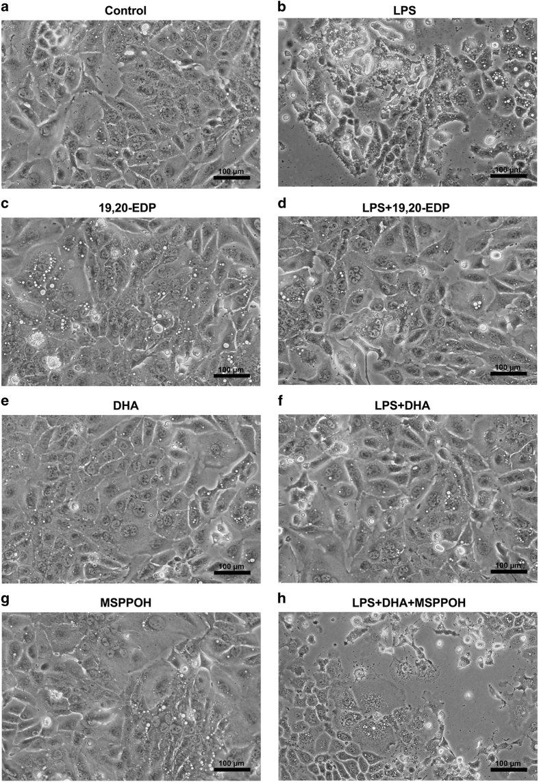
EDPs prevent LPS-induced morphological abnormalities in HL-1 cardiac cells. HL-1 cells were visualized by phase-contrast microscopy at 200× following treatment. (a) Untreated HL-1 cardiac cells. (b) HL-1 cardiac cells were stimulated with LPS (1 μg/ml) for 24 h. (c) HL-1 cells were treated with 19,20-EDP (1 μM) for 24 h. (d) HL-1 cells were treated with LPS (1 μg/ml) and 19,20-EDP (1 μM) for 24 h. (e) HL-1 cardiac cells were treated with DHA (100 μM) for 24 h. (f) HL-1 cardiac cells were stimulated with LPS (1 μg/ml) in the presence of DHA (100 μM) for 24 h. (g) HL-1 cardiac cells were treated with MSPPOH (50 μM) for 24 h. (h) HL-1 cardiac cells were stimulated with LPS (1 μg/ml) in the presence of DHA (100 μM) and MSPPOH (50 μM) for 24 h. Assessment of cell morphology was performed on a minimum of 15 cells per treatment. Scale bar, 100 μm in diameter.

