Figure 3.
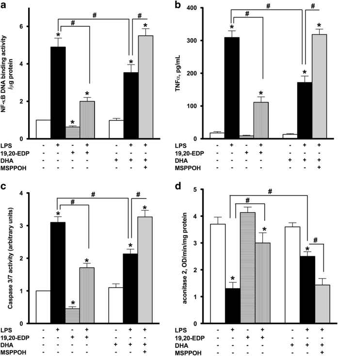
EDPs suppress LPS-induced inflammatory response, activation of caspase-3/7 and inhibition of aconitase 2. HL-1 cardiac cells were treated with LPS (1 μg/ml) in the presence of 19,20-EDP (1 μM), DHA (100 μM) and/or MSPPOH (50 μM) for 24 h. (a) NF-κB DNA-binding activity in the whole-cell lysates was measured by ELISA. (b) TNFα concentration in the culture supernatants was determined by ELISA. (c) Caspase-3/7 activity was measured in the whole-cell lysates by spectrofluorometric assay. (d) Aconitase 2 activity was measured in the whole-cell lysates by colorimetric assay. Values are represented as mean±S.E.M. N=3 independent experiments. *P<0.05 treatment versus vehicle control; # P<0.05 treatment group versus LPS or LPS/MSPPOH.

