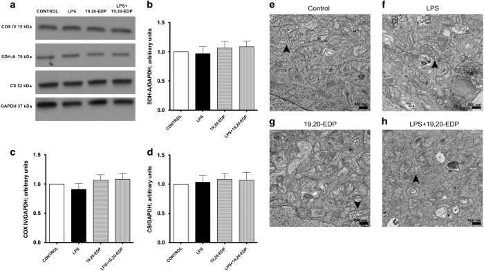
Figure 4.
EDPs limit LPS-induced damage to mitochondrial ultrastructure. (a) HL-1 cardiac cells were stimulated with LPS (1 μg/ml) with or without 19,20-EDP (1 μM) where indicated for 24 h. After 24 h, the whole-cell lysates were harvested and then analyzed by western immunoblotting for the levels of essential mitochondrial proteins. Representative western blots and the results of quantification are demonstrated in (a–d). HL-1 cardiac cells were treated as indicated above. Representative electron micrograph (EM) images of HL-1 cells are presented on (e–h). Black arrowheads demonstrate individual mitochondrion. Scale bar, 500 μm in diameter. Values are represented as mean±S.E.M. N=3 independent experiments.

