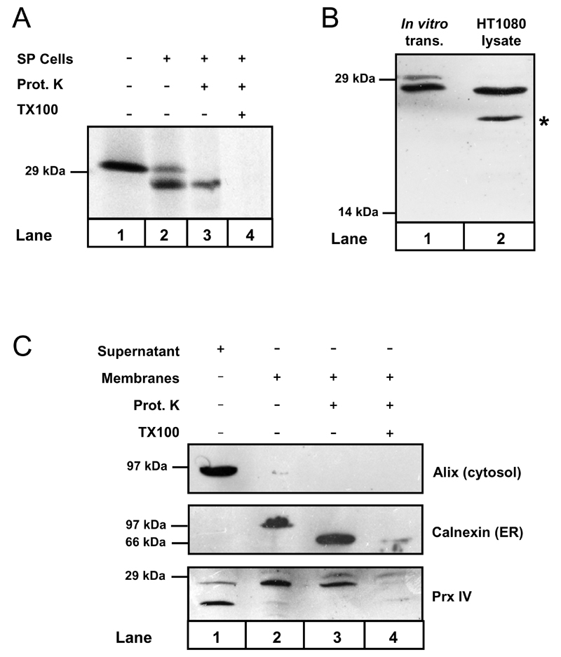
Figure 1. Prx IV is co-translationally translocated to the ER in vitro and in vivo.
A. Autoradiograph showing Prx IV mRNA translated in vitro using 35S-labelled methionine & cysteine. Translation was performed in the presence or absence of semi-permeabilised HT1080 cells (SP cells) as indicated. SP cells were harvested and treated with proteinase K with or without Triton X-100 detergent (TX100) as required. B. Prx IV was translated in vitro plus SP cells. Translation products were compared with HT1080 whole-cell lysate by Western blotting using antibody to Prx IV. C. HT1080 cells were homogenised and the post-nuclear supernatant separated by ultracentrifugation. The resulting supernatant and organelle membrane fractions were probed by Western blotting using antibodies to cytosolic and ER proteins along with anti-Prx IV. Membrane samples were also treated with proteinase K in the presence or absence of TX100 as indicated.

