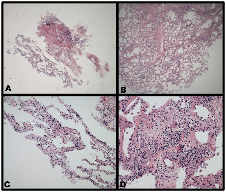
Figure 1. Representative pathology specimens.
(A) This low power demonstrates tissue adequacy obtained by TBLB. (B) This same power in the same patient demonstrates the relative amount of tissue obtained by TBLC. (C) This high power image of TBLB in the same patient demonstrates only non-specific inflammation. (D). This high power specimen obtained by TBLC in the same patient demonstrates ill-defined granulomas with a background of patchy lymphocytes and plasma cells.

