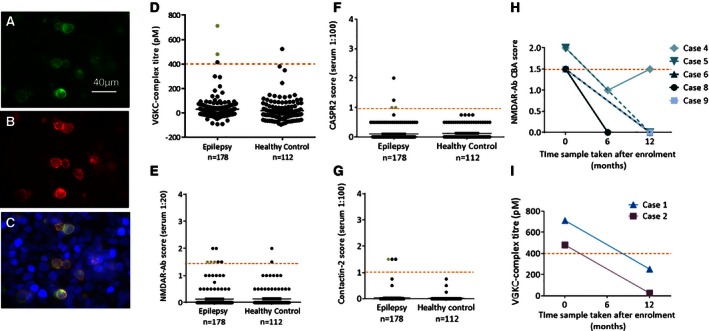
Figure 1.

Autoantibody testing results of the epilepsy and healthy control cohorts. The transfected CASPR2‐EGFP tagged transfected HEK cells (green) are shown (A). Serum from patient 16 binds to the surface of the CASPR2 transfected cells, seen with anti‐human IgG labelling (red, B).The transfected cells (A) and anti‐human immunoglobulin (IgG)–labelled cells (B) colocalize indicating a positive result for this patient (C). The scatter diagrams show the titers and CBA scores of positive tests for each antigen tested at the onset of epilepsy compared with healthy controls; VGKC complex (D), NMDAR (E), CASPR2 (F), and contactin‐2 (G). The red dashed line indicates the positive cut‐off used for each assay. The serum samples highlighted by green dots were positive on surface hippocampal staining in vitro. When available, follow‐up samples were tested; four of five NMDAR‐Ab positive patients and both VGKC‐complex antibody‐positive patients were negative at either 6 or 12 months after intake (H, I). Patient 4 showed an initial reduction in antibody levels then increase over time. These fluctuating antibody levels did not correlate with developmental regression or seizure activity.
