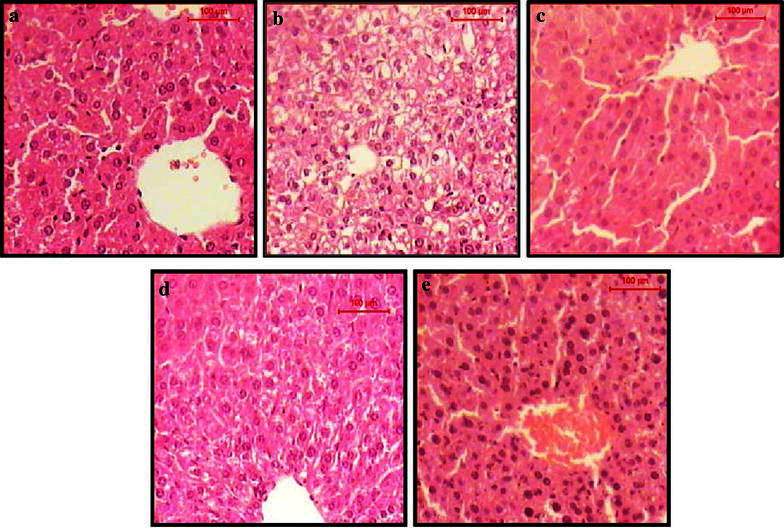
Fig. 3.

TS of liver of a normal control mice shows the normal architecture of hepatic lobule. The central vein surrounded by cords of hepatocytes. Between the strands of hepatocytes the narrow blood sinusoids are often seen, b EAC control mice shows hydropic degeneration of the hepatocytes, loss of cell boundaries and ballooning degeneration. Some other hepatocytes showed nuclear pyknosis and karyolysis, c 1/5 LD50 of venom-treated EAC-bearing mice shows the hepatic lobule that appear more or less like normal, d cisplatin-treated EAC-bearing mice shows the hepatic lobule that appear more or less like normal, some hepatocytes shows hydropic degeneration and ballooning degeneration, e 1/5 LD50 of venom, shows the hepatic lobule that appear more or less like normal. Notice the enlargement and hyperchromasia in the nuclei of the hepatocytes (H&E, scale bar 100 µm, magnification ×400)
