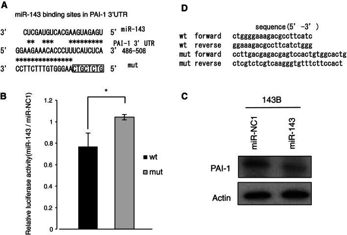
Figure 1.

PAI‐1 is target gene of miR‐143 in 143B cells. (A) Alignment of the wild‐type PAI‐1 3′UTR (wt) and mutant PAI‐1 3′‐UTR (mut) with the miR‐143 binding site, displayed in the 3′–5′ orientation. (B) Reporter assay for analysis of the luciferase activity of the wt or mut luciferase reporter in 143B after transient transfection of miR‐143. The data were normalized to the control miR‐NC1. *P < 0.05 (C) Western blot analysis of PAI‐1 expression in 143B after transient transfection of miR‐143 or the control miR‐NC1. PAI‐1 expression was quantified using ImageJ software and was normalized to β‐actin. Expression was calculated relative to that in miR‐NC1‐transfected cells. (D) The sequences of the wt and mut PAI‐1 3′UTR. PAI‐1, plasminogen activator inhibitor‐1; 3′UTR, 3′‐untranslated region.
