Figure 3.

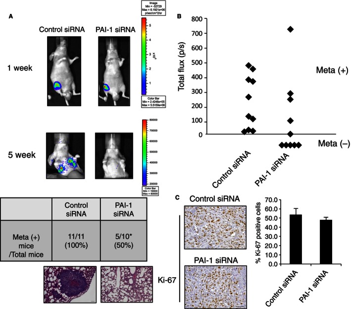
PAI‐1 siRNA regulates lung metastasis of 143B cells. (A) 143B‐luc cells were inoculated into the right knee of a model mouse. Representative bioluminescence at 1 and 5 weeks after inoculation as seen with the IVIS imager (top), and representative H&E staining of the lung at 5 weeks after inoculation (bottom) are shown. Scale bars, 100 μm. The table in (A) shows the number of mice with lung metastasis (meta +) at 5 weeks over the total number of mice in the two groups. (B) Total Flux (photons per second, p/sec) measured in the obtained IVIS images of mice‐transfected control siRNA or PAI‐1 siRNA lung metastasis at 5 weeks after siRNA inoculation. (C) Representative histochemical staining of Ki‐67 in primary tumor cells from control and PAI‐1 siRNA‐transfected mice. The percentage of tumor cells that were positive for Ki‐67 was calculated by counting 10 visual fields at high magnification. Scale bars, 50 μm. PAI‐1, plasminogen activator inhibitor‐1; IVIS, in vivo imaging system.
