Fig. 2.

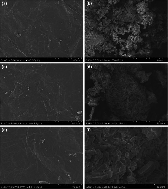
Scanning electron microscopy images of pristine GO and PEI-GO. The surface morphology of pristine GO (a, c, e) was compared with that of PEI-GO (b, d, f) by a JSM-6500 F SEM at different scales

Scanning electron microscopy images of pristine GO and PEI-GO. The surface morphology of pristine GO (a, c, e) was compared with that of PEI-GO (b, d, f) by a JSM-6500 F SEM at different scales