
Fig. 2.
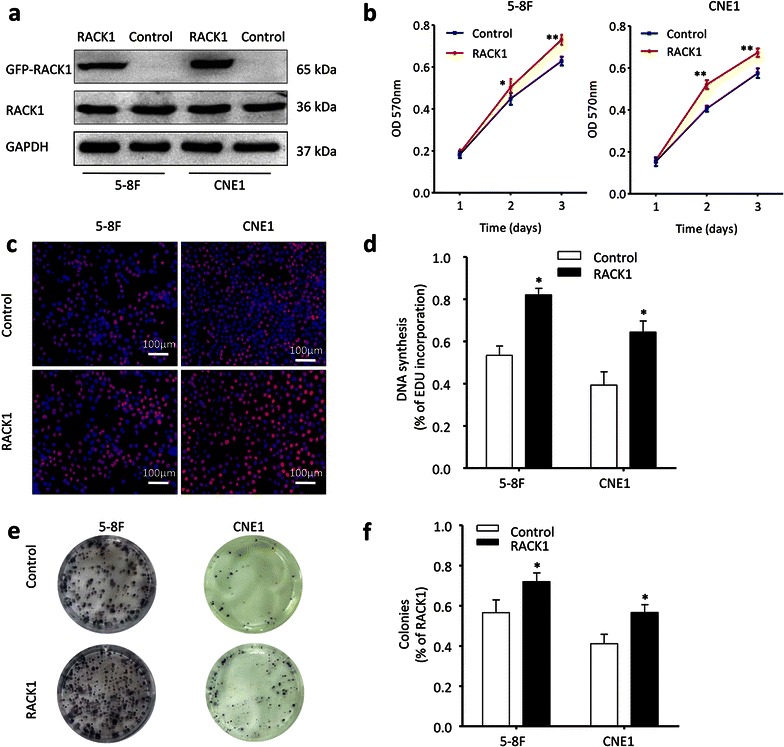
RACK1 propels NPC cells proliferation. a 5–8F and CNE1 NPC cells were transfected with GFP-RACK1 plasmid or control plasmid, and then analyzed by western blot. GAPDH served as the internal control. b MTT assays were conducted to detect the cell growth in indicated NPC cells. Two-way ANOVA test. c, d The cell proliferation status in NPC cells was also tested by EdU assay. Original magnification, ×100; scale bar 100 μm. Student’s t-test. e, f Besides, the clone formation ability of NPC cells was compared after transfected with RACK1 plasmid or control plasmid. Student’s t-test. Mean ± sem, N = 3, *P < 0.05, **P < 0.01, ***P < 0.001