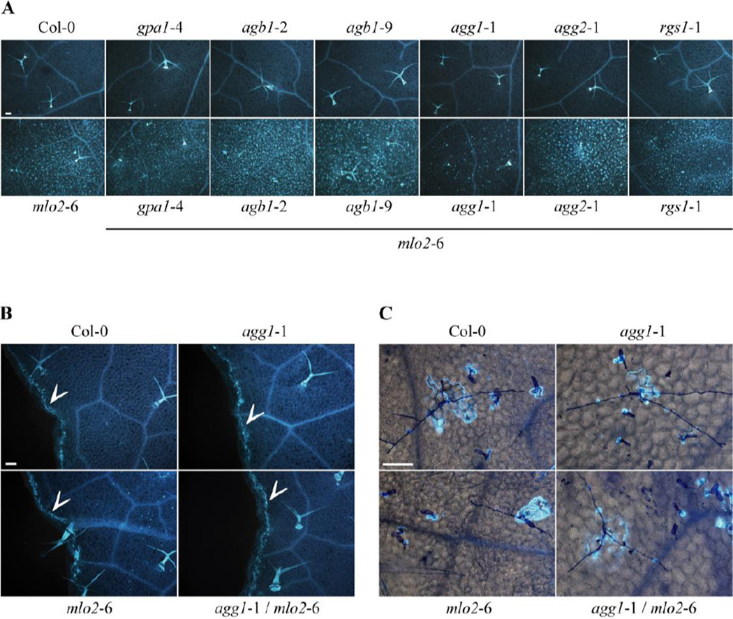
Figure 2.
Callose accumulation in rosette leaves. Callose was stained with aniline blue. (A) Representative micrographs demonstrating spontaneous callose deposition in leaves of 6-week old plants of the indicated genotypes grown in pathogen-free conditions. The experiment was repeated at least three times with similar results. Bar = 100 µm. (B) Leaves from 4-week old plants of the indicated genotypes injured with forceps showing callose deposition at wound sites (= arrowheads). The experiment was performed twice with similar results. Bar = 100 µm. (C) Four-week old leaves from plants of the indicated genotypes at 7 d post inoculation with the non-adapted powdery mildew fungus E. pisi exhibit callose deposition at sites of fungal interaction. Fungal structures were stained with Coomassie Brillant Blue. The experiment was performed once. Bar = 100 µm.

