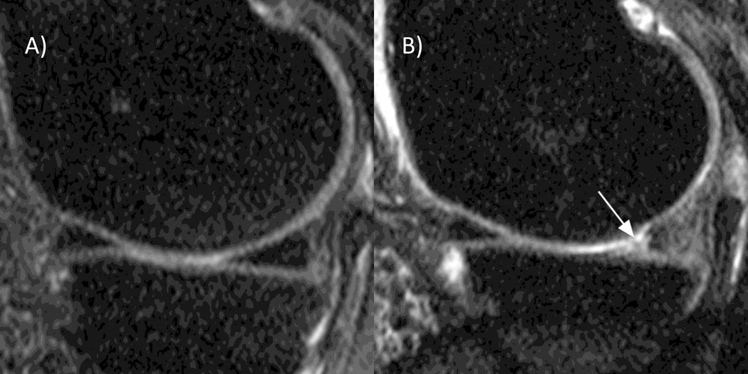
Figure 2. Meniscal tear.
A) Baseline sagittal fat-suppressed proton density-weighted 1.0T MRI shows normal triangular appearance of the posterior horn of the medial meniscus without tear or intramensical signal alterations. B) Follow-up image shows a meniscal tear reaching the superior and inferior surface of the posterior horn (arrow).

