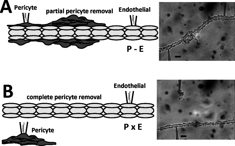
Fig 5. Configurations for simultaneous recording from DVR pericytes and endothelia.
A. Left and right panels show schematic depiction and photomicrograph of a partially denuded DVR with simultaneous dual-cell patch clamp of a pericyte and an endothelial cell that retain contact (abbreviated P-E). B. Left and right panels show a schematic depiction and photomicrograph of a DVR, fully denuded of pericytes, with simultaneous dual-cell patch clamp of each cell type when they have no contact (abbreviated PxE). These configurations were used to test the importance of cell contact for AngII dependent membrane potential responses. The black bars = 10 microns.

