
Fig. 5.
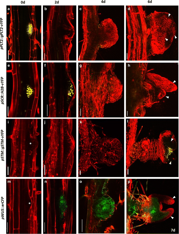
Confocal time lapse imaging during LRP to shoot conversion. a–d Time-lapse imaging showing PLT2-vYFP expression during the conversion of LRP to shoot. Note the down-regulation of PLT2-vYFP during the development of shoot meristem. e–h Dynamic expression pattern of pSCR::H2B-vYFP during the conversion of root to shoot. i–l Live imaging showing STM-vYFP expression during the shoot conversion. l Note the appearance of STM-vYFP in the nascent shoot meristem. m–p Spatiotemporal expression pattern of pWUS::erCFP. p Note the rapid upregulation of pWUS::erCFP upon cytokinin treatment. Arrowhead in (d, h, l, p) marks leaf primordium. Red signal is propidium iodide stain in (a, b, e, f, i, j, m, n) and FM4-64 stain in the remaining. Scale bar 50 µm