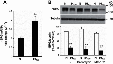
Fig. 3.

Effect of IH on hERG mRNA and the effects of proteosomal and lysomal inhibitors on IH-induced degradation of hERG protein. A: quantitative real-time PCR analysis of hERG mRNA levels in SH-SY5Y cells exposed to normoxia or IH120. The data are means ± SE from 4 individual experiments. B: representative immunoblot of hERG protein in cells exposed to normoxia and IH120 in the presence of baflomycin (1 μM) and MG-132 (5 μM), inhibitors of lysome and proteosome, respectively (top). Densitometric analysis of hERG protein normalized to tubulin expressed as percentage of normoxia (bottom). Data are means ± SE from 3 individual experiments. **P < 0.01, compared with normoxia.
