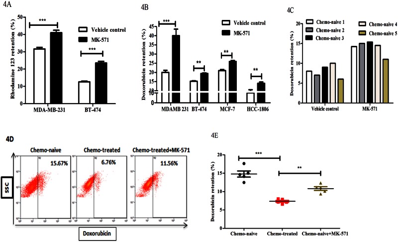
Fig 4. Knockdown of ABCC3 enhances doxorubicin retention in breast cancer cells.
A) Bar graph shows the rhodamine-123 retention (%) in MK-571 treated MDA-MB-231 (MFI-341.1±4) and BT-474 cells (324.1±0.6) and vehicle treated MDA-MB-231(MFI-302±2) and BT-474 cells (295±5) respectively. Error bar represent standard error of the mean (SEM); ** = p<0.01, *** = p<0.001, n = 3. B) Bar graphs shows the doxorubicin retention (%) in MK-571 treated MD-MB-231 (MFI-11±0.32), BT-474 (10.5±0.6), MCF-7 (MF-13.2±0.3) and HCC-1806 (MFI-5.9±0.5) and vehicle treated MD-MB-231 (MFI-15.5±0.3), BT-474 (8.3±0.9), MCF-7 (12.1±0.2) and HCC-1806 (MFI-4.1±0.2) breast cancer cell lines respectively. C) Bar graphs shows the doxorubicin retention (%) in MK-571 treated chemo-naive tumour derived cells (MFI-14.5±0.8) compared to vehicle treated cells (MFI-9.7±0.5). Error bar represent standard error of the mean (SEM); ** = p<0.01, *** = p<0.001, n = 3. D and E) FACS plots (4D) shows the doxorubicin retention in chemo-naive tumour derived cells (n = 5, MFI-9.8±0.45), CT treated tumour derived cells (n = 5, MFI-6.1±0.3) and CT treated tumour derived cells treated with MK-571 (MFI-7.7±0.4). Scatter plot shows the doxorubicin (%) retention in tumour derived cells (4E). Error bar represent standard error of the mean (SEM); n = 3.

