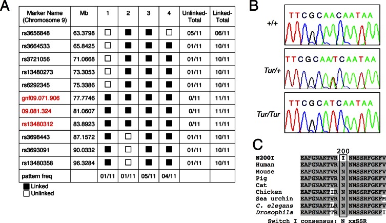
Fig 2. The Turner mutation in myosin VI.
(A) Pattern program output of the results of 11 markers located between 63.3798 Mb and 96.3284 Mb, tested on DNA derived from 11 affected (9 were G3 offspring and 2 were the N2 breeding pair), that showed recombination between three markers (gnf09.071.906, 09.081.324 and rs13480312). The Tur mutation was determined to lie between marker rs6292345 (75.3386 Mb) and rs3698443 (87.1572 Mb). The black box (linked) indicates mice heterozygous for the C3H and the C57; the white box (unlinked) indicates mice homozygous for the C3H (patterns 1 and 2) or the C57 (pattern 4). (B) Sequence analysis of the Turner genomic DNA revealing a 820A>T transversion in exon 8 (NM_001039546). Control DNA shows a single A peak and heterozygous DNA (Tur/+) shows both A and T peaks, but homozygous DNA (Tur/Tur) shows a single T peak. (C) The p. N200I single amino acid substitution. Alignment of a portion of myosin VI protein from various species revealing conservation of the N200 residue. The switch I loop consensus sequence motif within the motor domain is indicated.

