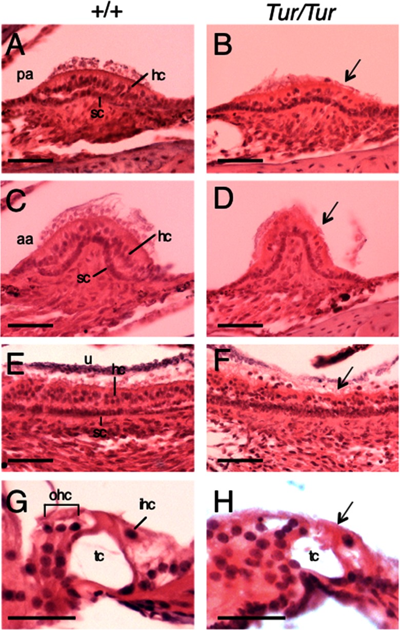
Fig 3. Degeneration of vestibular hair cells in Tur/Tur mutant.

H&E staining of inner ear tissue at 4 weeks. (A-H) Posterior (A,B) and anterior (C,D) crista ampullaris, utricular macula (E,F) and Organ of Corti (G,H) in wild-type (A,C,E,G) and Tur/Tur mutant (B,D,F,H). Significant reduction of hair cells in the vestibular sensory organs (arrows in B,D,F) and collapse of the organ of Corti (arrow, H) are evident in the inner ear of Tur/Tur mice. Panels G,H are images showing the organ of Corti in the basal turn. Abb.: hc, hair cell; ihc, inner hair cell; ohc, outer hair cell; sc, supporting cell; tc, tunnel of Corti. Scale bar: A-F, 50 μm and G,H, 25 μm.
