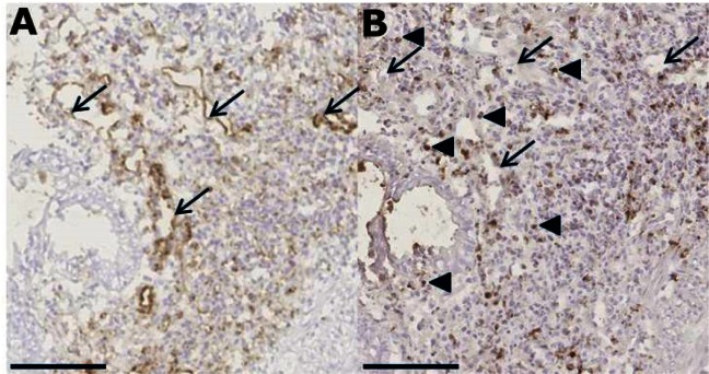
Fig 4. Antibodies in the culture medium diffuse into the biopsy and bind to target cells.
Inflamed colonic biopsies were cultured in the presence of anti-CD31 and anti-CD3 antibodies (10 μg/ml each) for 20 hours followed by IHC staining of consecutive sections with isotype-specific anti-IgG2a (A) and anti-IgG1 (B) antibodies for detection of anti-CD31 and anti-CD3 antibodies, respectively. Arrows indicate blood vessels, which were stained in (A) by the anti-IgG2a secondary antibody specifically recognizing the anti-CD31 antibody. Blood vessels were not stained by the anti-IgG1 secondary antibody (B) that instead recognized the binding of the anti-CD3 antibody to T cells in the lamina propria (indicated by arrowheads). Bar = 100 μm.

