
Fig 5. LPS uptake is mediated by the LDLR in human hepatocytes.
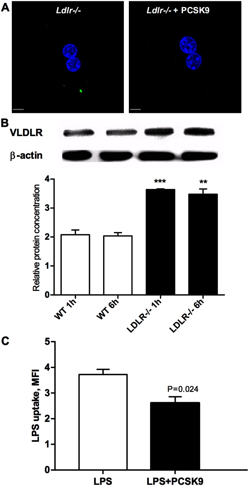
(A) PCSK9 further reduced LPS uptake in Ldlr-/- cells. Primary hepatocytes were isolated from Ldlr-/- mice. Cells were allowed to attach to coverslips for 6–12 hours than treated with 2.5 ug/mL Alexa Fluor 488–conjugated LPS for 6 hours total. Coverslips were fixed with 4% paraformaldehyde for 20 min and stained with DAPI to identify nuclei (blue). LPS uptake was visualized using Leica Inverted Fluorescence microscope (x63 magnification) and analyzed with VELOCITY software. Images are representative of three independent experiments. (B) Ldlr-/- mice have increased expression of VLDLR in a liver. VLDR receptor protein expression by Western blot in wild type and Ldlr-/- mouse liver at time points 1 hour and 6 hours after injection of LPS. Data presented as mean±SEM, (***P = 0.0007, compared with control 1h; **P = 0.0026, compared with control 6h, n = 3). (C) PCSK9 further altered uptake of LPS in HepG2 cells with LDLR knockdown. HepG2 cells were reversely transfected with siRNA targeting LDLR and control scrambled siRNA. Recombinant human PCSK9 (3 μg/ml) was added 4 hours before and 4 and 19 hours after LPS treatment. Cells were treated with Alexa Fluor 488-conjugated LPS (2.5 μg/ml) 48 hours after transfection for 24 hours. LPS uptake was measured by flow cytometry. Data presented as mean fluorescence intensity, mean±SEM (n = 3).