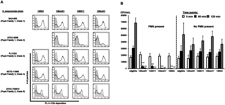
Fig 2. Anti-PspA mAbs show activity in complement deposition and opsonophagocytic killing assays.
(A) Pneumococci were incubated with 0.25–5 μg/mL mouse isotype control or anti-PspA IgG2a mAbs and mouse serum. Bound C3 was detected with a FITC-labeled anti-mouse C3 antibody using flow cytometry. Histograms: FL-1 for isotype (tinted; gray lines) or the respective anti-PspA (black lines) mAbs. Note: 139G3 did not bind to ATCC-6305. (B) S. pneumoniae PJ-1324 cells were pre-opsonized with 1 μg/mL mouse IgG2a control or anti-PspA mAbs in 10% baby rabbit complement and 106 PMN-like HL-60 cells or vehicle. At the indicated times, CFU were enumerated. Samples were run in triplicate or quadruplicate and mean values + SD of one representative experiment of several performed are shown (***, p<0.0005; anti-PspA vs. isotype mAb, unpaired t-test).

