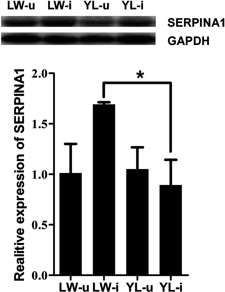
Fig 8. Changes in SERPINA1 protein expression after PCV2 infection.
Western blotting was used to measure SERPINA1 protein expression in each group of lung tissue samples. The SERPINA1 level in LW-i was significantly higher than that in YL-i. GAPDH expression was used as the positive control. * P < 0.05.

