
FIGURE 5:
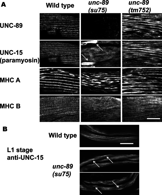
Localization of thick filament proteins in the body wall muscle of wild type and two categories of unc-89 mutants. (A) Adult muscle. Each box contains an immunofluorescence image of portions of several body wall muscle cells reacted with antibodies to UNC-89, paramyosin, or one of the two myosin heavy chains (MHC A or MHC B). unc-89(su75) lacks all giant UNC-89 isoforms (Small et al., 2004), and thus the paramyosin-binding SH3 domain is missing. unc-89(tm752) is an unc-89 mutant in which all of the kinase-containing isoforms are missing but giant isoforms (UNC-89-A and -E) are present (Ferrara et al., 2005). The antibody used to detect UNC-89, EU30, was developed to Ig domains 3–6 and thus detects the giant isoforms (Small et al., 2004). Note the large accumulations of paramyosin in unc-89(su75), indicated by arrows. Although unc-89(tm752) shows disorganization of UNC-89, paramyosin, and MHC A, no accumulations of paramyosin are detected. Therefore, when the paramyosin-binding SH3 domain is missing, paramyosin forms large accumulations outside the normal thick filaments. (B) L1 larval-stage muscle reacted with anti-paramyosin. In wild-type animals, paramyosin is localized to A-bands, each body wall muscle cell having two A-bands. In unc-89(su75), abnormal accumulations of paramyosin are seen, indicted by arrows (examples of two animals). Scale bars, 10 μm.