FIGURE 3:
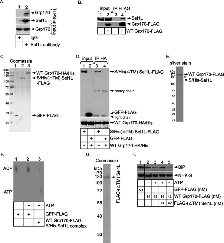
Enzymatically active Grp170 binds to the Hrd1 adapter Sel1L. (A) Cells were incubated with the DSP cross-linker (1 mM) for 30 min, quenched, and semipermeabilized with digitonin, and the resulting pellet fraction was treated with Triton X-100. The resulting lysate was incubated with either a control immunoglobulin G or Sel1L-specific antibody, followed by protein G–agarose beads. The precipitated samples were analyzed by SDS–PAGE and immunoblotted with the indicated antibodies. (B) WCEs derived from Flp-In T-Rex-293 parental cells and cells stably expressing WT Grp170-FLAG were subjected to precipitation using FLAG antibody–conjugated agarose beads. The isolated proteins were analyzed by SDS–PAGE and immunoblotted with the indicated antibodies. Input WCEs were also analyzed by immunoblotting with the indicated antibodies. (C) Coomassie staining of purified C-terminally FLAG-tagged GFP (GFP-FLAG), C-terminally HA- and His-tagged WT Grp170 (WT Grp170-HA/His), and N-terminally S-/His-tagged, C-terminally FLAG-tagged transmembrane-deleted Sel1L [S/His(ΔTM) Sel1L-FLAG]. (D) WT Grp170-HA/His (150 nM) was incubated with either S/His(ΔTM) Sel1L-FLAG (150 nM) or GFP-FLAG (150 nM), and the samples were immunoprecipitated using an anti-HA antibody. The immunoprecipitates were subjected to SDS–PAGE, followed by immunoblotting with an anti-FLAG antibody. The input samples were also analyzed by immunoblotting with an anti-HA antibody. (E) Silver stain of the WT Grp170-FLAG:S/His-Sel1L complex. Asterisk denotes degraded WT Grp170-FLAG. (F) FLAG-BiP was incubated with [α-32P]ATP to form the radiolabeled ADP–BiP complex. The ADP–BiP complex was then incubated with GFP-FLAG or the isolated Grp170–Sel1L complex indicated in the absence or presence of unlabeled ATP. ADP release form BiP was analyzed by TLC. (G) Coomassie staining of purified, N-terminally FLAG-tagged, transmembrane-deleted Sel1L (FLAG-(ΔTM)Sel1L). (H) As in Figure 2B, except that the Grp170-FLAG was preincubated with or without FLAG-(ΔTM)Sel1L at 37ºC for 30 min before being added to the BiP-NHK-S complex. The black line indicates that intervening lanes in the same immunoblot have been spliced out.

