
Figure 1.
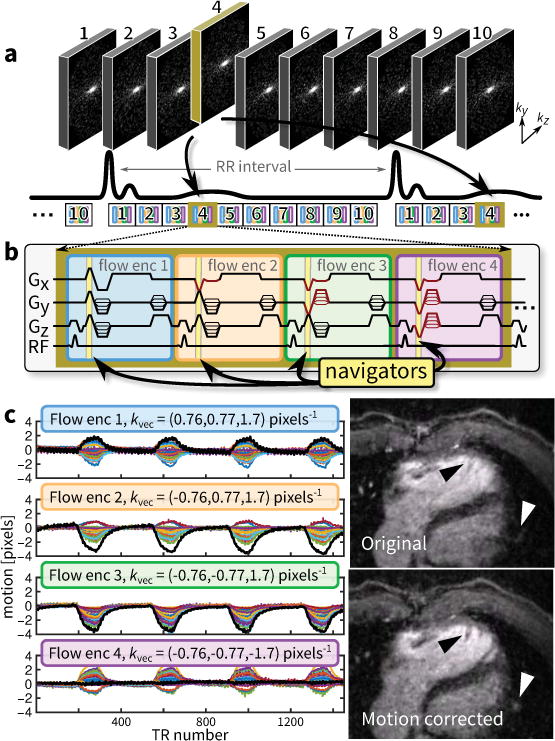
Data acquisition overview for 4D flow. a: Sampling and view-ordering of 10 cardiac phases determined using VDRad that is aligned with the cardiac triggering – for longer RR intervals, patterns are repeated before the next RR interval. b: One flow-encoding unit consisting of four different flow-encoding configurations with built-in Butterfly navigators acquired during the flow-encoding gradients. c: Motion estimated (cardiac motion suppressed with a stop-band filter) from different flow-encodings – each color is from a different channel in a 32-channel cardiac coil receiver. In c, the uncorrected and corrected diastolic phase images (10% acceptance window) from a 4D flow scan using VDRad (9.1 min, R = 23) of a 22-year-old male is shown. To demonstrate the accuracy of the motion estimates, the image is corrected using the linear motion estimate with the largest range (thicker black line in c). The right ventricular trabeculae are sharpened (black triangle); peripheral pulmonary vessels are recovered (white triangle). Because the motion is non-rigid, other portions of the image are blurred. For this work, the motion estimate with the largest range is used to compute weights for the image reconstruction.