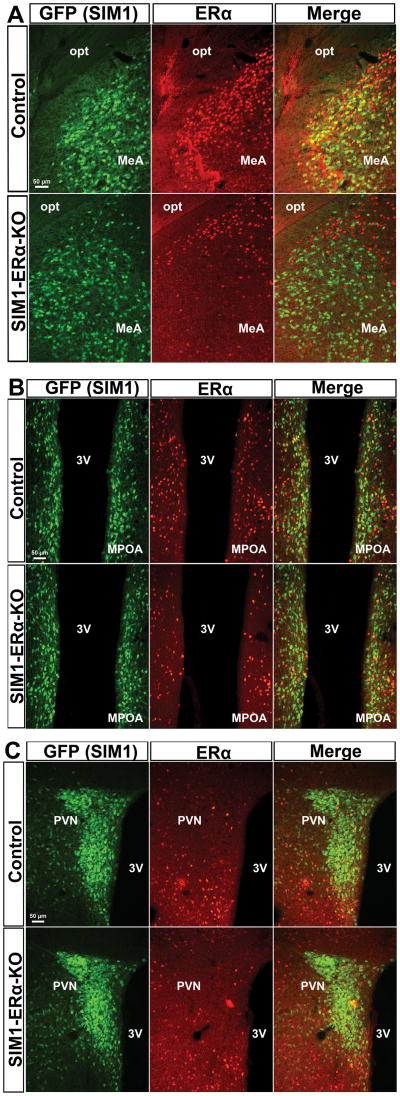
Figure 1. Deletions of ERα in SIM1 neurons.
Dual immunofluorescence for GFP (green) and ERα (red) in the MeA (A), MPOA (B) and PVN (C) in control (SIM1-Cre/Rosa26-GFP, upper panel) and SIM1-ERα-KO (Esr1f/f/SIM1-Cre/Rosa26-GFP mice, lower panel) mice. 3V, third ventricle; MPOA, medial pre-optic area; opt, optic tract. Scale bars = 50 μm.

