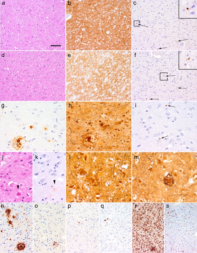
Fig. 1.
Neuropathology of iCJD cases. Mild to moderate spongiform change in the HE staining (a, d) associated with diffuse/synaptic PrP immunoreactivity (b, e) and focal tau immunoreactive neuritic profiles (c, f indicated by arrows, a representative one is enlarged in right upper inset) in case iCJD-1 (a–c) and iCJD-2 (d–f). The same mature plaque with a corona in iCJD-2 as seen in immunostaining for Aβ (g), Bielschowsky (h), AT8 (i arrows indicate small neuritic profiles as seen in CJD cases but not around the mature plaque), HE (j arrowhead) and Congo staining (k arrowhead indicates the plaque as seen under polarized light). Bielschowsky staining (l, m) of two mature plaques lacking tau immunoreactivity close to each other in iCJD-2. Immunostaining for Aβ (n) and ubiquitin (o) in the same cortical regions close to the dura transplant in iCJD-1. Immunostaining for AβPP in frontal white matter in iCJD-1 (p) and iCJD-2 (q). Immunostaining for HLA-DR (microgliosis) in the frontal cortex (r) and white matter (s) in iCJD-1. The bar in image “a” represents 100 μm for a–f, r, s; 40 μm for g–m; and 60 μm for n–q

