Fig. 2.
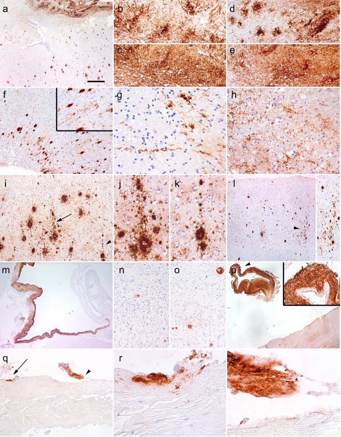
Aβ immunoreactivity in iCJD-1. Immunostaining using the 6F/3D Aβ antibody (a, b, f, g, i–m, p–s), the 4G8 Aβ antibody (c, h), anti-Aβ1–40 (e, n), and anti-Aβ1–42 (d, o) demonstrated widespread immunoreactivity in the lesion area (a upper part enlarged in b–e); radiating deposits in the cortex (f) and white matter (g, h); focal deposits with columnar alignment perpendicular to the surface of the cortex (i–l); in a dilated vein (m), as immunostaining of plaques (n, o); and as dural deposits (p–s). The right upper inset in p is the enlargement of the area indicated by an arrowhead; r and s are enlargements of areas indicated by arrow and arrowhead in q, respectively. The bar in image “a” represents 150 μm for a, f, l, m–o; 60 μm for b–e; 40 μm for g, h, j, k; 100 μm for i, p, q and 15 μm for r, s

