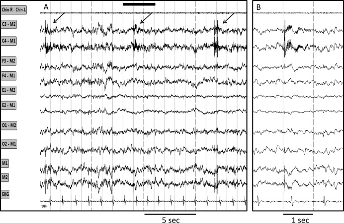
Figure 6. Tracings from one epoch showing intermittent very high frequency bursts limited to the central electroencephalograph (EEG) derivations.
(A) 20-sec epoch. (B) Faster tracings in the region outlined by the solid bar in A. Chin R-Chin L, chin electromyogram; C3, C4, F3, F4, O1, O2, M1, M2 are electroencephalography tracings from left and right central, frontal, occipital and mastoid electrodes; E1 and E2, left and right eye electrodes. EKG, electrocardiogram.

